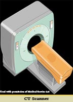
 Cardiac CT (“CAT scan”) is a non-invasive technique which uses concentrated radiation directed at the beating heart to produce a three-dimensional picture of the heart and surrounding blood vessels. The patient is placed on a moving table which enters the “doughnut” which spins an X-ray camera around the patient’s chest at high speed. The test is painless and typically claustrophobic patients do not have difficulty.
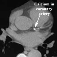
In a non-contrast study, CT can screen for calcium which is a surrogate for cholesterol plaques. This technique gives a rough estimate as to the amount of total cholesterol affecting the arteries that feed the heart. It cannot determine the severity of blockage in individual arteries nor necessarily determine the cause of chest pain. In this type of scan, a low score indicates a very low risk for heart attack and a high calcium score predicts a high overall risk which should be treated with aggressive control of blood pressure, cholesterol and blood sugar (if needed).
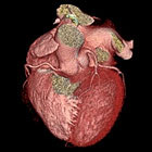
 A contrast-enhanced study provides a much more detailed image of the heart. With this type of exam the size of the main chambers and blood vessels can be determined as well as the overall function of the heart. Most of the heart’s blood vessels can also be visualized in fine detail and the severity of any blockages can be determined. Unfortunately, this technique is not very good at looking through stents or at very small blood vessels in which case coronary angiography is needed. In addition, there is limited amount of information provided in patients who have undergone bypass surgery in the past as the metal clips used to attach the grafts interfered with proper visualization of the artery.
This type of exam is not for every patient as there are risks involved. Those who should not have the exam include pregnant women, patients with an allergy to contrast and those with kidney disease. In addition there is a significant amount of radiation exposure equivalent to approximately 100 chest X-rays or more. Your physician will help you decide whether or not this test is right for you.
 Some insurance plans will not pay for this procedure so the hospital will check to make sure that this test is best for you.
Patient preparation for the exam consists of avoiding any food or liquids 4-6 hours prior to the exam and to remove any metal objects such as jewelry which may interfere with the images obtained. An IV is placed to administer contrast. If the heart rate is too rapid, a medication called a beta-blocker(or calcium channel blocker) will be administered to slow the heart rate in order to obtain higher resolution pictures. During the exam the patient is placed on a table which slides through a doughnut shaped scanner. The exam itself only takes 5 to 10 minutes. After the exam you will be instructed to drink plenty of liquids to flush out the contrast, but otherwise you may immediately resume normal activities.
To learn more about Cardiac CT, click here.