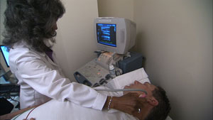
Carotid ultrasound is a technique using ultrasound waves to look for atherosclerotic blockages in the two of the main arteries feeding the brain. Similar to what is used to image an unborn baby in pregnant women, this process is safe and does not require IV’s or other more invasive techniques. Since these arteries are located in the neck close to the skin, high fidelity pictures can be directly obtained of the artery and the blood which flows through them.
 By studying these images, we are able to assess if there is any obstruction to the blood flow through these arteries. Significant obstructions can be the cause of a stroke or TIA (transient ischemic attack), often called a “mini-stroke.” Symptoms are often manifested as sudden changes in speech or vision as well as numbness or weakness affecting one side of the body.
From a patient perspective, there is no preparation for the exam necessary and all medications can be taken as usual. A gel will be applied to the neck to help conduct the ultrasound waves more efficiently. The procedure takes approximately 15 to 30 minutes. After scanning the arteries, the gel is wiped off and the patient may then leave the facility. Once the study is processed and interpreted, the results will be communicated to the ordering physician.
To learn more about Carotid Ultrasound, click here.